12Sledge and double sledge microtome
around 1930
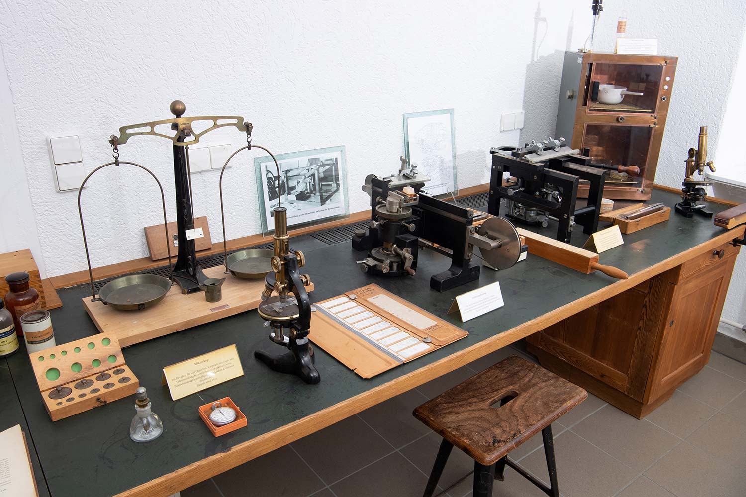
Workstations like this one were used for histological examinations: studies of the shape or anatomy of cells or tissues and how they change in cases such as disease. Such anatomical assessments require examining the tissue under the microscope. This requires cutting it into very thin sections so that light can penetrate the object, which is necessary in transmission light microscopy.
It is impossible to make tissue sections that are thin enough by hand – they need to be thinner than a human hair and evenly preserve tissue structure. Thus, to make the sections, scientists use a device called a microtome. In one type, a sledge microtome, the knife is guided through the tissue along a sled. Double-sledge microtomes were used to produce tissue sections with uniform thicknesses from larger specimens such as entire organs. Here, the blade is guided through the object by being held in two sleds.
This makes precision cutting devices such as the microtomes exhibited here, from the company R. Jung, Heidelberg, indispensable for histology. Without them, it would have been impossible for early neuroscientists such as Oskar and Cécile Vogt and Korbinian Brodmann to systematically analyze brains and determine the types of cell and tissues they found.
Ultramicrotomes can make even thinner slices. One such instrument is on display next to the electron microscope.
You can find more information about the production of permanent histological specimens in the video with this title, on the website of the virtual microscope museum.
You can find more information about the life and work of Oskar and Cécile Vogt here.